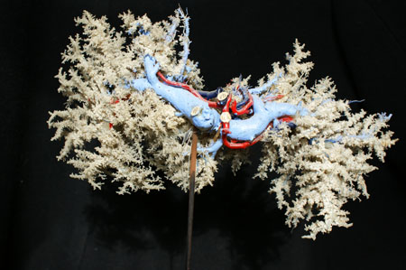
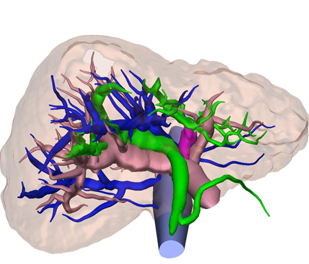
Corrosion preparation of liver vessels. Preparation was made by Nechunaev L.M., 1960.
CATEGORY SCIENTIFIC
![]()
Corrosion preparation of liver vessels. Preparation was made by Nechunaev L.M., 1960.
![]()
Corrosion preparation of liver vessels. Preparation was made by Nechunaev L.M., 1960.
![]()
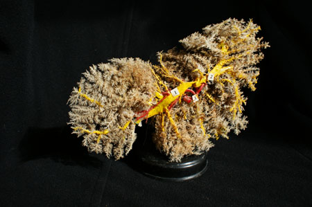
Corrosion preparation of liver vessels. Preparation was made by student
Miftahov S., under the guidance of Gladkih A.V., 1954.

![]()
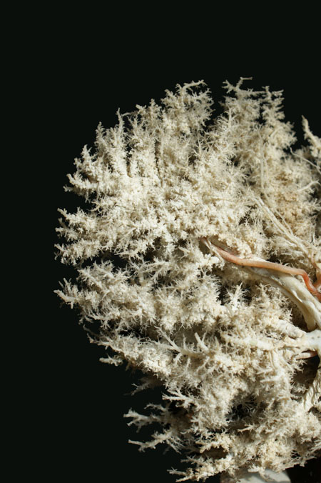
Corrosion preparation of liver vessels. Preparation was made by student Miftahov S.,
under the guidance of Gladkih A.V., 1954.
![]()
Corrosion preparation of liver vessels. Preparation was made by student
Miftahov S., under the guidance of Gladkih A.V., 1954.
![]()
HSC.
![]()
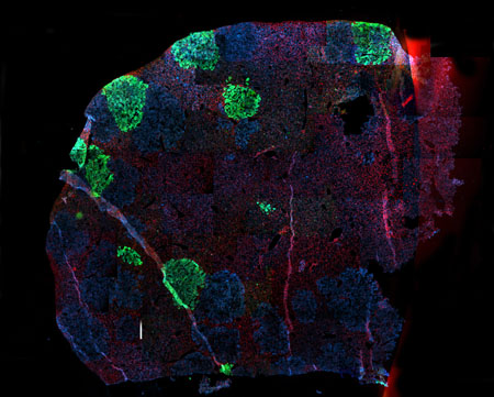
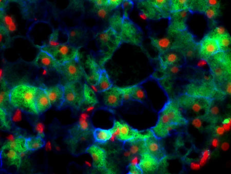
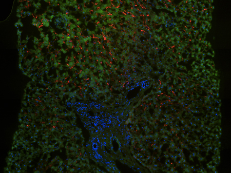
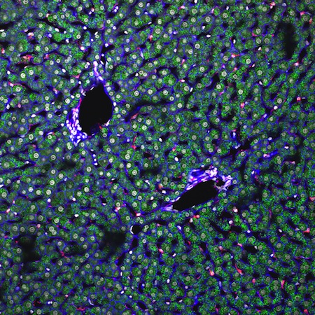
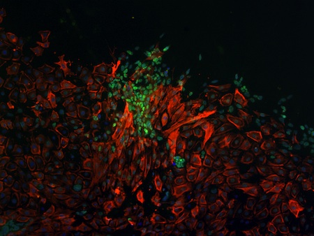
uPA mice were transplanted with GFP-expressing (green) or native
human hepatocytes (orange staining for human albumin) and engraftment
was monitored 2 months after transplantation. This image has been sampled
together using 64 overlays (3-fluorescence channels, blue: nucleus).

![]()
uPA mice were transplanted with GFP-expressing (green) or native
human
hepatocytes (orange staining for human albumin) and engraftment
was monitored 2 months after transplantation. This image has been sampled
together using 64 overlays (3-fluorescence channels, blue: nucleus),
false colour image.
![]()
uPA mice were transplanted with human hepatocytes co-expressing cytoplasmic
GFP (green) and murine CD47. This image shows a false colour overlay of a human
hepatocytes cluster with strong transmembrane expression of murine CD47(blue)
while murine hepatocytes only demonstrate weak endogenous expression of
mCD47 (red: nucleus).
![]()
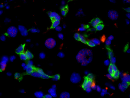
uPA mice were transplanted with human hepatocytes expressing GFP (green)
and or not. Red cell nucleus staining (false colour) visualizes the separate clusters
of human hepatocytes that have engrafted. Human albumin (blue) is synthesised
within the cells and secreted, explaining the blue staining in the intercellular space.

![]()
uPA mice were transplanted with human hepatocytes co-expressing cytoplasmic
GFP (green) and murine CD47. This image shows a false colour overlay of a human
hepatocytes cluster with strong transmembrane expression of murine CD47(blue)
while murine hepatocytes only demonstrate weak endogenous expression of
mCD47 (red: nucleus).
![]()
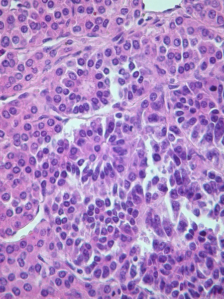
Hepatocellular carcinoma arising from hepatocellular adenoma
with intrahepatic metastasis.

![]()
Ground glass hepatocytes.

![]()
ALPHA -1 ANTITRYPSIN GLOBULES IN HEPATOCYTES - PAS staining.

![]()
ALPHA -1 ANTITRYPSIN GLOBULES IN HEPATOCYTES - PAS staining.

![]()
ALPHA -1 ANTITRYPSIN GLOBULES IN HEPATOCYTES - H&E staining.

![]()
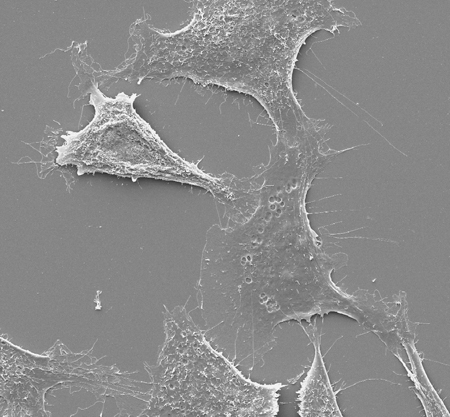
Explantation growth of hepatic cells from newborn rat`s liver, 48h, x20.

![]()
Explantation growth of hepatic cells form newborn rat`s liver, 48h, x20.

Liver - Anatomic approach.

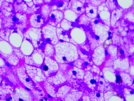
Liver biopsy, may grunwald giemsa stain, liver steatosis.

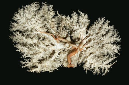
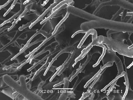
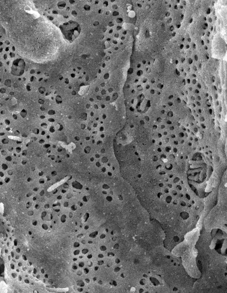
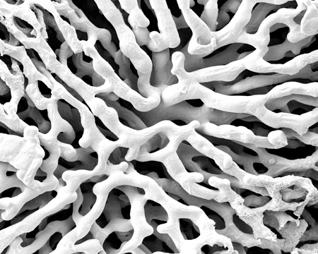
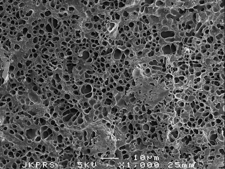
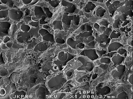
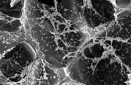
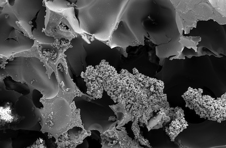
EM photography of a cast of the liver microvasculature.
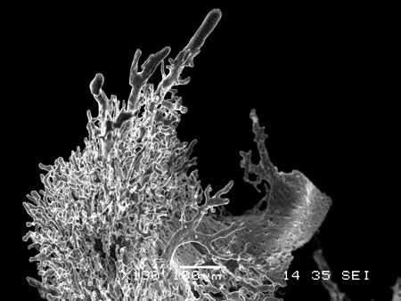
EM photography of a cast of the liver microvasculature.
EM photography of a cast of the liver microvasculature.

EM photography of a cast of the liver microvasculature.
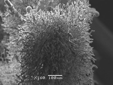
EM photography of a cast of the liver microvasculature.
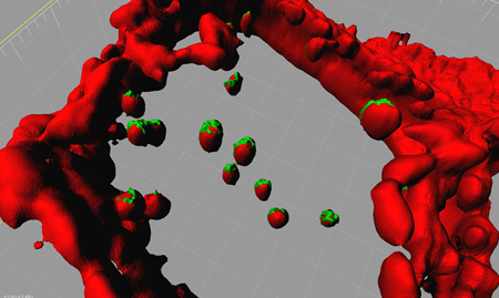
Liver, partial hepatectomy with hepatic stellate transplantation, 2nd day,
EGFP+ transplanted cells, x40.

Culture of hepatic stellate cells, 1st passage, 22nd day, cells in mitosis,
immunostaining for desmin - marker of rat`s hepatic stellate cells, x40.

Co-culture of Hepatic stellate cells (green, PKH67 labeled) and Bone Marrow
derived mesenchymal Stem Cells (red, PKH26 labeled) on matrigel, 1st day, x10).

Immunohistochemistry assay for desmin-positive hepatic stellate cells in the liver after
partical hepatectomy and cultivated hepatic stellate cells transplantation, 7th day, x20 .
Hepatic veins viewed from the right atrial.
Neurofibrillary proteins in Liver.
Second Harmonic Generation SHG (left) and simultaneous TPEF/SHG (right)
acquisitions of non-stained fibrillar collagen deposit in human cirrhotic liver.
Extracellular matrix reconstruction of SHG images of non-stained collagen
in human fibrotic liver.

Talks between cells (TPEF images).

HCV direct cell-to-cell transmission. Naïve Huh7.5-RFP-NLS-IPS cells (Red)
surrounding Jc1-infected Huh7.5-EGFP-IPS cells (green) become infected during
co-cultivation in the presence of a neutralizing antibody that blocks cell-free infectivity.

3D-imaging simulation using a method for fusion of CT and MRCP images.
3D-imaging simulation using a method for fusion of CT and MRCP images.

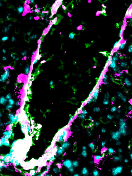
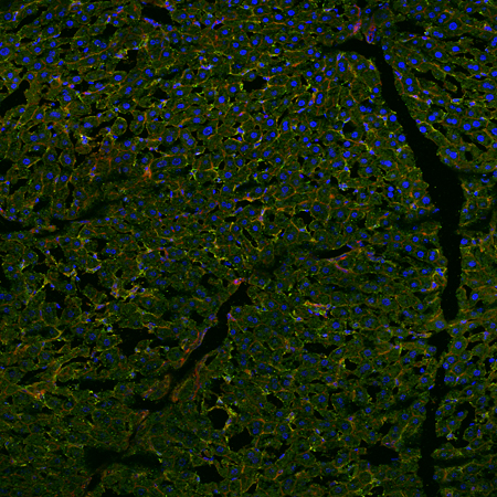
Topological distribution of HLA-class I viral peptide complexes on HBV infected liver.
Red is HLA-class I/HBV envelope peptide complexes, green is cytokeratin, Blue is Dapi.
Comment: Presentation of HLA-class I/ viral peptide complexes is detectable throughout
the liver parenchyma.
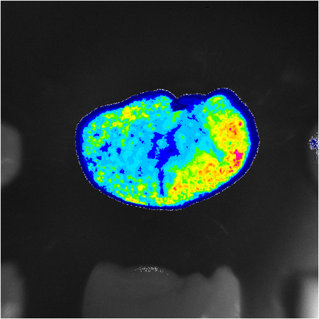
Whole liver luciferase imaging from fasted (left) and fed (right) ERE-Luc transgenic mice.
The increased light on the right liver is because of a mTOR-dependent ER activation by
dietary amino acids (Della Torre, Rando et al., Cell Metabolism 2011 13(2) 205-14).

Dietary activation of the estrogen receptor in mouse liver. Luciferase imaging
magnification of the left hepatic lobe, note the zonation of ER activity
(Della Torre, Rando et al., Cell Metabolism 2011 13(2) 205-14 ).
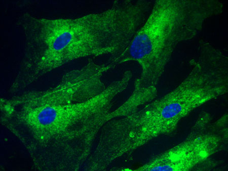
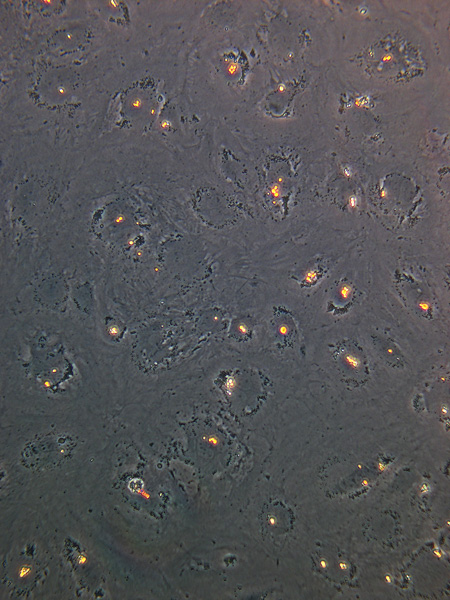
Glycogen depots (PAS staining) in mouse primary hepatocyte cultures.

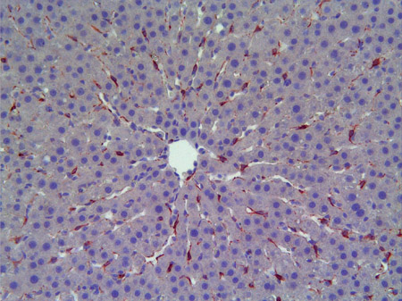
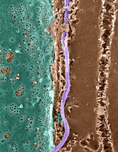
Pseudo-colored Scanning EM of an hepatic plate, accentuating the fenstrated sinuoidal
endothelium (blue), heptaocyte (brown) and a collagen fibril (lavender).
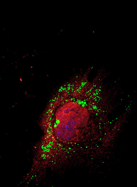
Isolated rat hepatocytes subjected to hypoxia-reoxygenation stained for Factor-inhibiting
hypoxia-inducible factor (red), which colocalizes with the peroxisomal marker PMP70
(green) and
not the mitochondrial marker ATP Synthase (blue). Colocalization of
red and green yields yellow.
DAPI-stained nuclei are pseudo-colored purple.
Confocal immunofluorescence microscopy.

Scanning electron micrograph of hepatocyte microvilli protruding through the sinusoidal
endothelial cell fenestrations.
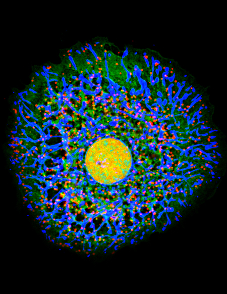
Isolated GFP-transgenic rat hepatocyte stained for peroxisomes (red),
mitochondria (blue) and
nucleus (yellow-orange).
Confocal immunofluorescence microscopy.
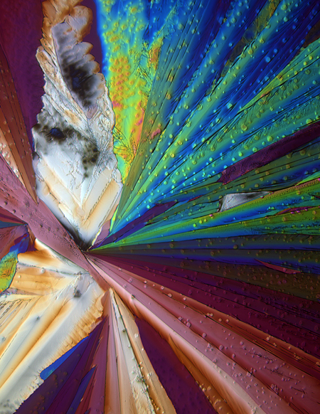
Polarized light image of acetominophen recrystalized on a slide.
Desmoglein 2 (red) & gamma-catenin (green) at the desmosomes in the liver.
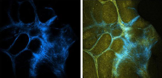
SEM of bile canaliculi of a liver with conditionally deleted beta-catenin.

Kupffer cells in vitro from normal human liver specimens.
ErbB2 expression of the mouse gallbladder carcinoma.
ErbB2 expression in the mouse primary gallbladder carcinoma.
Ultrasound image of mouse gallbladder carcinoma.

Actin-dependent phagocytosis in liver professional phagocytic cells.
Confocal laser scanning microscopy and reconstitution using IMARIS software.
Up taken particles (green) are surrounded by actin (red) phagocytic cup structures.
Gram-negative bacteria and liver infections - Electron microscopy.

Scanning electron micrograph of isolated murine liver sinusoidal endothelial cells.
Scanning electron micrograph of an isolated murine liver sinusoidal endothelial
cell exhibiting fenestration.

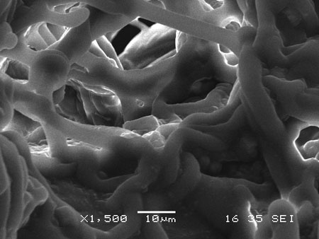
Scanning electron micrograph of a vascular corrosion cast depicting the sinusoidal
network in a mouse liver.
Scanning electron micrograph of a vascular corrosion cast illustrating the complex
network of hepatic sinusoids in a mouse liver.

Scanning electron micrograph showing fenestrated sinusoids and hepatocytes
in a mouse liver.

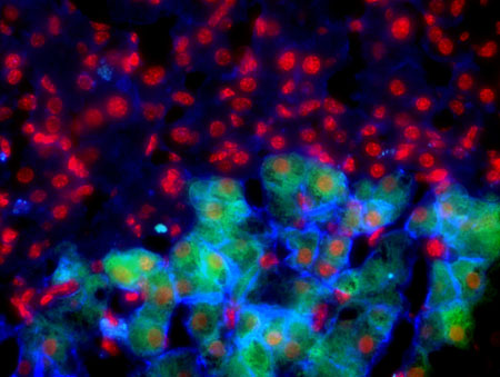
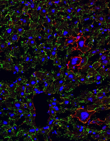
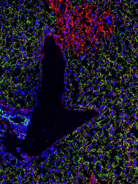
Experimental model of spontaneous chronic liver injury. One essential mediator
of immune response (MyD88) was inactivated in mice to investigate the role of
this pathway in the development of chronic liver disease. DAPI (blue) shows
all the cell nuclei; CK-19 (green) stains putative progenitor cells; SOX9 (red)
differentiating hepatocytes after liver injury (400 µm).
Immunofluorescent staining using antibody against HEV ORF2 antigen in primary
human hepatocyte. (x400 - 1week after infection)

Immunofluorescent staining using antibody against HEV ORF2 antigen in
human
hepatocyte. (x400 - 2week after infection)
Immunofluorescent staining using antibody against HEV ORF2 antigen in human
hepatocyt.e (x630 - 3week after infection)
Immunofluorescent staining using antibody against HEV ORF2 antigen in
primary human hepatocyte. (x400 3week after infection) The infected cells were
found to form “infected cell clusters” and the number of the clusters rather stayed
constant. However, the number of infected cells in each cluster was found to increase
with passage of culture time.This finding suggests that HEV infection spreads over
by cell-to-cell transmission of virion in human hepatocytes.
Immunofluorescent staining using antibody against HEV ORF2 antigen
in primary human hepatocyte. (x200 3week after HEV infection).

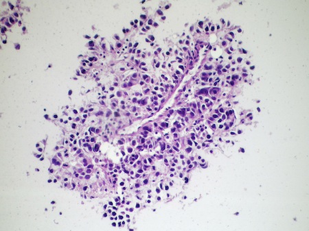
Neoplastic proliferation of small cells showing an
organoid arrangement with
prominent vascular pattern. The neuroendocrine
lineage was confirmed by
inmunohistochemistry. (FNA cell-block, H&E x 100)
In embryonal hepatoblastoma, small, elongated embryonal cells form pseudo rosettes.
Scanning electron microscope image (1000x) from normal human liver sample
Scanning electron microscope image (1000x) from cirrhotic human liver sample
Vit A crystals in hepatic stellate cell..
Autophagy and in vitro hepatic stellate cell activation
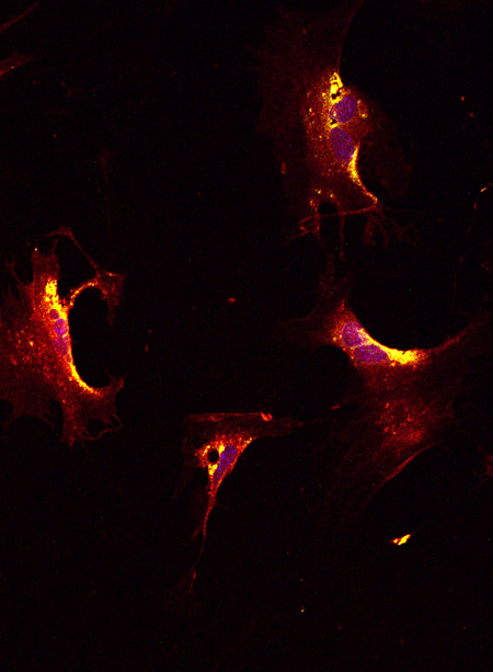
Autophagy during hepatic stellate cell activation (LC3B and lipid droplet co-localization)
Stellate cells and Vit A lipid droplets.

Chemoembolisation

Medical procedure

Gamma-catenin compensates for conditionally deleted beta-catenin
at adherens junctions
Gamma-catenin compensates for conditionally deleted beta-catenin
at adherens junctions
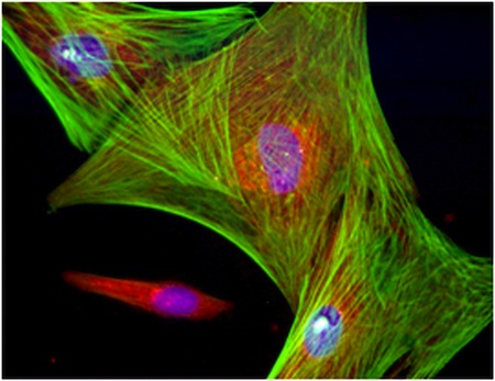
Rat Liver. 20x magnification. White is dapi nuclear stain, green is
Laminin, red is alpha tubulin and blue is 647 phalloidin actin marker.
Rat Liver section, 20x magnification, white is Dapi nuclear stain,
green is pmp70, red is alpha tubulin and blue is 647 phalloidin actin marker.
Rat Liver. 40x mag with a 1.3x digital zoom factor. White is dapi
nuclear stain, green is PMP70, Red is smooth muscle actin and
blue is
647 phalloidin actin marker.
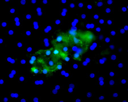
Different population of non-parenchymal liver cells in culture

Liver progenitor cells in culture
Liver progenitor cells in culture surrounded by other
non-parenchymal cells
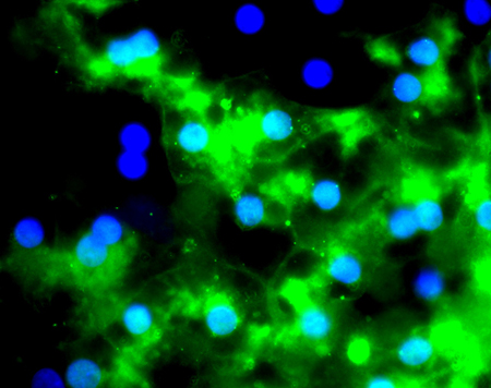
Liver progenitor cells in culture expressing Alpha Fetal Protein
and Cytokeratin

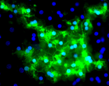
Liver Progenitor cells in culture with non-parenchymal cell population.

SEM of Hepatic stellate cells on PDLLA Scaffold (x100).
SEM of Hepatic stellate cells on PDLLA Scaffold (x50).

SEM of Hepatocytes on PDLLA Scaffold (x50).
Histological patterns in drug-induced liver disease.
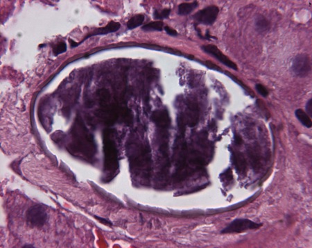
Hepatic biopsy. Images compatible with Schistosoma Japonicum ova.
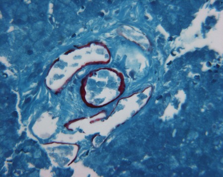
Hepatic biopsy. Images compatible with Schistosoma Japonicum ova.

Hepatic biopsy of 49-yrs old woman from Philippines, in Italy for the last 20 yrs.
Hepatic biopsy shows images compatible with Schistosoma Japonicum ova.
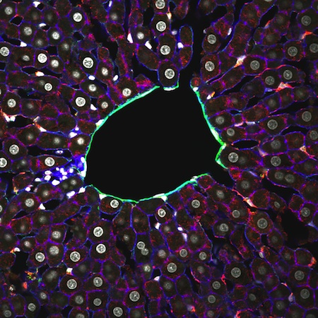
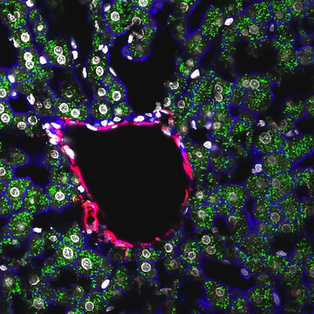
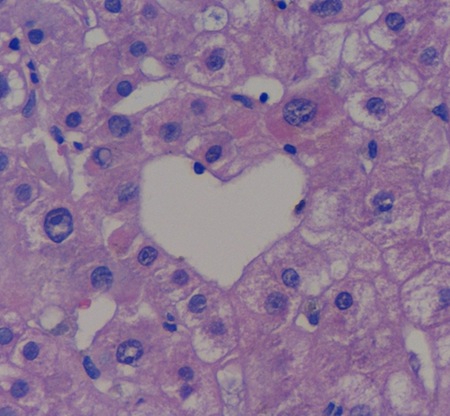
"Liver heart" - A heart-shaped terminal hepatic venule in a liver biopsy,
Haematoxylin-eosin, x40
Alpha-1-antitrypsin globules in a transgenic mouse model
for alpha-1-antitrypsin deficiency (diastase PAS reaction).

No description submitted